Surgical treatment of varicose veins of the lower limbs began in the modern era approximately 100 years ago with the work of Mayo and Babcock. Even today, the basic principle remains the same. In recent years, surgical results have been significantly improved by three main innovations: improved surgical technique based on new insights into venous anatomy and pathophysiology, the routine use of preoperative color Doppler ultrasound, and the adoption of less invasive surgical techniques (miniphlebectomy and invagination stripping).
The frequency of varicose vein operations in Europe varies considerably — from 70 procedures per 100,000 inhabitants in the United Kingdom to 200 per 100,000 in Finland, up to 100,000 operations per year in Italy and as many as 150,000 per year in France. The goal of these procedures is to relieve symptoms and prevent complications.
The objective of surgical therapy is the elimination of superficial reflux in the saphenous vein trunk, the interruption of hydrodynamic forces caused by reflux in insufficient perforating veins, and the removal of all visible varicose veins.
WHEN DO WE DECIDE ON SURGICAL TREATMENT OF VARICOSE VEINS?
Surgical treatment is indicated in the presence of:
- Unsightly appearance
- Pain
- Heavy legs
- Rapid fatigue of the lower limbs
- Recurrent superficial thrombophlebitis
- Bleeding from ruptured varicose veins
- Hyperpigmentation around the ankle
- Lipodermatosclerosis
- Atrophie blanche
- Venous leg ulcer
Surgical Treatment Options
- Removal of the saphenous vein from the groin to the ankle with additional phlebectomies
- Segmental removal of the saphenous vein with additional phlebectomies
- Ligation of the saphenous vein (high, mid, or low) with sclerotherapy
- Ligation of the saphenous vein with additional phlebectomies
- Phlebectomies alone
- Endovenous laser closure of the diseased long or short saphenous vein
New Surgical Techniques
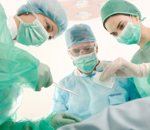
The classical operation for varicose veins — high ligation of the great saphenous vein (GSV) at its junction with the femoral vein, stripping of the GSV in the thigh, and removal of as many varicose tributaries as possible — also has unwanted side effects. These include hematomas, infections, damage to cutaneous sensory nerves, and potential complications of spinal or general anesthesia.
Furthermore, this type of treatment requires hospitalization and a relatively long period of absence from work. In an effort to eliminate the need for anesthesia, hospitalization, and prolonged sick leave, several less invasive methods for treating varicose veins have been developed: sclerotherapy, ultrasound-guided and catheter-directed sclerotherapy, local miniphlebectomy, and radiofrequency closure of the GSV.
All these methods have highly variable success rates, which depend primarily on how effectively they can achieve the goals mentioned above. Other modern minimally invasive techniques use heat generated by the impedance of radiofrequency (RF) energy or the thermal effect of a laser beam to obliterate (close) the GSV in the immediate vicinity of the saphenofemoral junction (SFJ). Early reports on RF closure of the GSV described success rates of 90–95% (after an average of 4.9 months), but also reported complications resulting from the effect of heat on the skin.
Endoscopic Subfascial Surgery of Perforating Veins (SEPS)
It has been known for nearly a century that insufficient calf perforating veins are associated with the most severe stages of chronic venous insufficiency (CVI). The classical operation described by Linton in 1938 was abandoned due to a 24% complication rate caused by poor surgical wound healing. Although many authors recommended modifications to reduce complications, it was only with the introduction of endoscopic subfascial perforator surgery that a significant reduction in complications was achieved.
In nine published studies involving a total of 465 limbs, only a 5% wound complication rate was reported. Ulcer healing was achieved in 90% of cases, with a 12% recurrence rate of leg ulcers. These results are good when compared to the usual success rates of leg ulcer treatment (50% recurrence), and therefore justify the use of this technique in patients with CVI stages C2–C6 who have insufficient perforating veins confirmed by color Doppler ultrasound.
Despite the favorable effect of the operation on ulcer healing and reduction of recurrent leg ulcers, there is still doubt regarding its overall effectiveness due to the lack of comparative studies between classical superficial varicose vein surgery with saphenous vein removal and SEPS. To date, no reports in the known studies have evaluated the impact of SEPS on quality of life after surgery.
At the General Hospital Novo mesto, since 1996, we have been performing operations on patients with CVI stages C2–C6 in which the classical varicose vein operation (high ligation at the saphenofemoral junction and removal of the GSV trunk in the thigh) is supplemented with endoscopic subfascial ligation of perforating veins (SEPS).
The operation is performed under tourniquet (bloodless field). It begins with the removal of the affected superficial veins and the GSV trunk. A 2–3 cm incision is then made in the skin and fascia, and an endoscopic instrument for subfascial ligation of perforating veins is introduced under the fascia. This instrument has one working channel that allows the use of endoscopic tools up to 7 mm in diameter and a standard connection for the endoscope and video camera. Using the instrument, we perform subfascial dissection distally to a few centimeters above the medial ankle. Perforators appear as vertical structures running from the muscles toward the fascia. We apply 5 mm metal clips to the perforators and divide them between the clips.